. 2018 May 29;9(25):5630–5639. doi: 10.1039/c8sc01288a

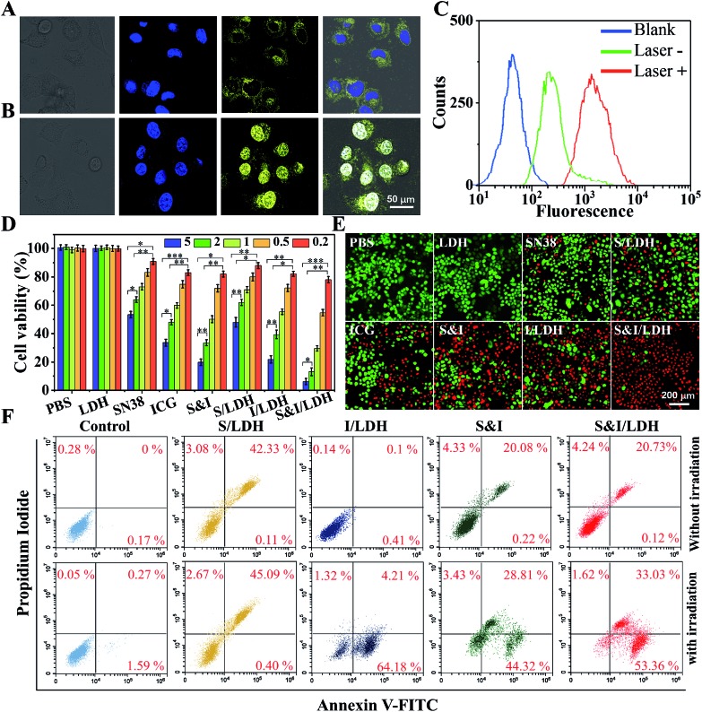
Fig. 4. Confocal images of Hela cells incubated with SN38&ICG/Gd&Yb-LDH: (A) before and (B) after NIR laser irradiation (808 nm, 1.0 W cm–2). (C) Intracellular fluorescence with and without NIR irradiation recorded using a flow cytometer. (D) Viability of Hela cells in the presence of the drugs with different concentrations (from 0.2 to 5.0 μg mL–1) upon NIR irradiation: (1) PBS, (2) Gd&Yb-LDH, (3) SN38, (4) ICG, (5) SN38&ICG, (6) SN38/Gd&Yb-LDH, (7) ICG/Gd&Yb-LDH, and (8) SN38&ICG/Gd&Yb-LDH and (E) the corresponding calcein-AM/PI staining images (5.0 μg mL–1). (F) Cell apoptosis analysis using the PI/annexin V–FITC double staining method. P values in (D) were calculated by Tukey's post-test (***P < 0.001, **P < 0.01, or *P < 0.05).

Fig. 4