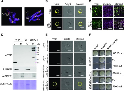
Figure 6.
OsPM1 Is Located at the Plasma Membrane.
(A) The secondary structure of OsPM1 predicted by PyMOL software (http://robetta.bakerlab.org/queue.jsp).
(B) Subcellular localization of OsPM1 in rice protoplasts. Bars = 20 μm.
(C) Subcellular localization of OsPM1 in transgenic rice plants. Young roots from 10-d-old rice seedlings expressing YFP-OsPM1 or YFP were observed. The fluorescence signal of FM4-64 indicates the plasma membrane. Bars = 50 μm.
(D) Immunoblot analysis of leaf cell lysates from Arabidopsis plants expressing YFP-OsPM1 and YFP. C, cytoplasm; M, plasma membrane. Antibody names are indicated on the left. Antitubulin and anti-PIP2;7 antibodies were used to verify the purity of cytoplasm and the plasma membrane portions, respectively. Coomassie blue staining of the SDS-PAGE gel was included as loading control.
(E) BiFC assay showing that OsPM1 interacts with itself in rice protoplasts. Coexpression of nYFP and cYFP-OsPM1, cYFP and nYFP-OsPM1, and an unrelated membrane protein control, nYFP-OsPIP2;4 and cYFP-OsPM1, were used as the negative controls for nYFP-OsPM1 and cYFP-OsPM1 coexpression in rice protoplasts. Bars = 20 μm.
(F) mbSUS assay showing that OsPM1 interacts with itself in yeast. Wild-type Nub (NubWT) and the mutant Nub (NubG) vectors served as the positive and negative controls of NubG-OsPM1, respectively. CubPLV acted as the negative control for OsPM1-CubPLV. FD indicates SD/-Ade, -His, -Trp, -Leu, -Met. Bars = 1 mm.

