Fig. 1.
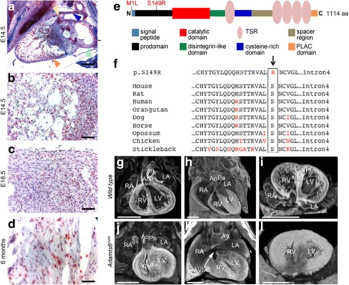
Adamts6 cardiac expression, sequence conservation, and cardiac anomalies in Adamts6-deficient mice. a–d Adamts6 (red punctate signal) is expressed in the outflow tract (a, blue arrowhead), heart valves (a, yellow arrowhead), atria (a, green arrowhead), and ventricular myocardium (a, orange arrowhead, b-d). e, f Diagram of the two Adamts6 mutant alleles recovered: Met1Ile and Ser149Arg. The sequence alignment shows conservation of the Ser149 residue in ADAMTS6 across species. g–l Congenital heart defects observed in Adamts6 Ser149Arg (Adamts6m/m) mutant embryos. A WT mouse heart with normal atrial, ventricular, and outflow tract anatomy (g), an intact atrioventricular septum (d), and normal ventricular myocardium (i). Homozygous Adamts6 Ser149Arg mutants (Adamts6m/m) exhibit a spectrum of congenital heart defects, such as a double outlet right ventricle (j, in which the aorta and pulmonary artery both arise from the right ventricle; see Additional file 3: Video S1) or an atrioventricular septal defect (AVSD) (k, in which the atrial and ventricular septa fail to form). Thickening of the ventricular wall is commonly observed, indicating ventricular hypertrophy (l). These mutant hearts (j–l) are shown at embryonic day (E)16.5 but their development is delayed, giving an appearance similar to WT hearts at E14.5 (as shown in (g–i)). Ao aorta, AVSD atrioventricular septal defect, LA left atrium, LV left ventricle, Pa pulmonary artery, RA right atrium, RV right ventricle. Scale bar: (a) 500 μm; (b–d) 50 μm; (g–l) 1 mm

