
Figure 1. DGKζ and Cbl-b deficient T Cells have enhanced ERK1/2 and IκBα phosphorylation.
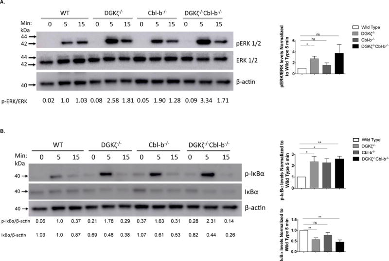
4×10⁵ STEMCELL-purified WT, DGKζ−/−, Cbl-b−/−, or DKO naïve CD8+ T cells were incubated with biotinylated α–CD3 (5ug/ml) and α–CD28 (5ug/ml) and cross-linked with streptavidin (25ug/ml) for the indicated times. Lysates were immunoblotted for protein levels of (A) phosphorylated ERK (p-ERK), total ERK (tERK) and β-actin or (B) phosphorylated IκBα (p-IκBα), total IκBα (IκBα) and β-actin. Relative band intensities are indicated below each lane. Representative blots are depicted from one of three independent iterations and ImageJ quantification of the three iterations are displayed ± SEM in the graphs.