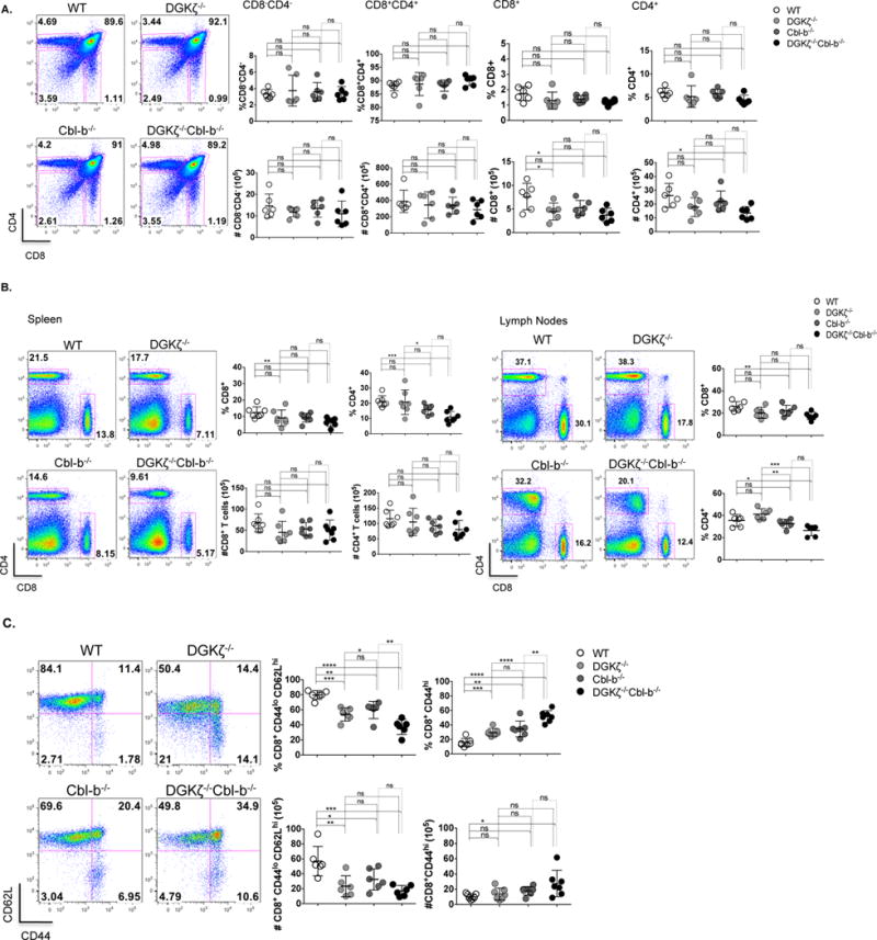
Figure 2. Loss of DGKζ and Cbl-b results in a greater percentage of splenic CD8+ T cells with an activated phenotype.

(A) Thymocytes, (B) splenocytes and lymph node cells from WT and knockout mice were stained for cell surface markers CD8 and CD4. Percentage and absolute number were determined for different cell populations. (C), Splenocytes were stained for cell surface markers CD8 and CD4, and CD62L and CD44 after gating for CD8+ cells, and the percentage and absolute number of CD8+CD44hi cells was determined. (n=7 for each group).
