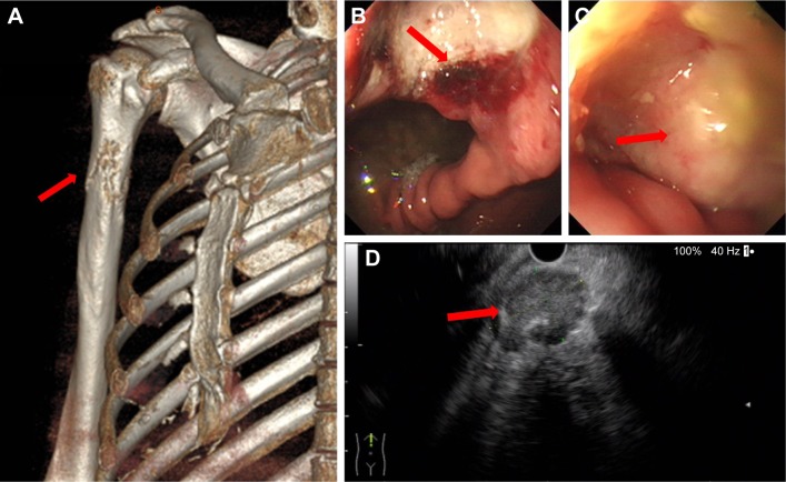
Figure 2.
(A) Three-dimensional reconstruction of computed tomography image revealed that the right upper humeral bone metastasis was combined with a pathological bone fracture (arrow). (B) Gastroscopy revealed an ulcer (arrow) of approximately 2×2 cm located in posterior wall of gastric corpus. (C) A rough uplift (arrow) of 1.5×2.0 cm was observed in the junction of duodenal bulb and descending part. (D) Endoscopic ultrasound-guided fine needle aspirate was performed on mediastinal lymph nodes (arrow).

