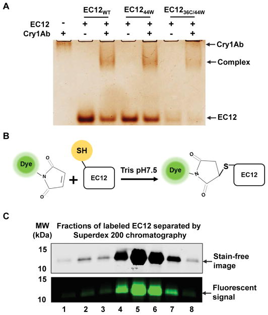
Figure 3.
Fluorescent labeling of mutant EC12 peptide.
(A) EC12WT and mutant EC12 show similar binding capacity to Cry1Ab. Binding reactions of the EC12 peptides to Cry1Ab were conducted at room temperature for 15 min. Indicated samples were resolved on a native gel and silver stained afterwards.
(B) Reaction showing the irreversible labeling of the cysteine residue on EC1236C/44W. “Dye” refers to Alexa-488, a fluorescent dye, which is conjugated to the maleimide moiety. Maleimide crosslinks the free thiol group depicted on the EC12 peptide.
(C) Separation of Alexa-488 labeled EC1236C/44W from free dye molecules by size exclusion chromatography. The peak protein fractions were resolved by SDS-PAGE. Stain-free images (top panel) were captured using a ChemiDoc MP Imaging system (Bio-Rad) and fluorescent images (bottom panel) recorded using a Typhoon FLA 9500 fluorescent scanner (GE Healthcare Life Science).

