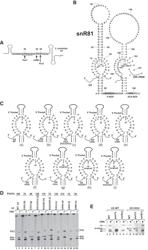
FIGURE 2.
Effects of mutations of U2 snRNA on Ψ42 formation within U2 snRNA. (A) Schematic representation of S. cerevisiae U2. The sequence of branch site recognition region, including the branch site recognition motif (underlined), is shown. The three pseudouridines, Ψ35, Ψ42, and Ψ44, and their respective enzymes, Pus7, snR81, and Pus1, are indicated. Pus7 and Pus1 are stand-alone proteins, and snR81 is a box H/ACA RNP. (B) Sequence and structure of snR81 box H/ACA RNA. The primary sequences of snR81 and its substrates, U42 (indicated by the arrow) of U2 (5′ pocket) and U1051 (indicated by the arrow) of 25S rRNA (3′ pocket), are shown. The H and ACA boxes are shaded. (C) Base-pairing interactions between the snR81 5′ pocket and U2. Depicted are base-pairing interactions between the guide sequence of the 5′ pocket and its U2 substrate sequences, either wild-type (a) or mutants (b–i). Italicized letters represent mutated nucleotides. Crosses (Xs) indicate disrupted base-pairing interactions. In (g–i), the original U42 is also indicated. (D) Detection of Ψs in yeast U2 using CMC-modification followed by primer-extension. Wild-type U2 (lanes 1 and 2) and various mutant (lanes 3–20) U2 snRNAs (illustrated in C, a–i) were assayed for pseudouridylation. Signals corresponding to Ψ35, Ψ42, and Ψ44 are indicated. Shifted Ψ42 bands are indicated by asterisks (*). The formation of Ψ42 was quantified using the formula Ψ42/(Ψ35 + Ψ42 + Ψ44). The mutants (mutant lanes) were then normalized to the wild-type control. The final pseudouridylation efficiency numbers are shown at the top of each lane. Note, in lane 12, although there is still some formation of Ψ42 at the original position, pseudouridylation is mostly shifted to the 5′-most uridine site. (E) U42-to-Ψ42 conversion by snR81. Pseudouridylation of wild-type U2 (lanes 1–6) and C41U mutant U2 (lanes 7–12) was analyzed in wild-type (lanes 1 and 2, and lanes 7 and 8), pus7-deletion (lanes 3 and 4, and lanes 9 and 10), and snr81-deletion (lanes 5 and 6, and lanes 11 and 12) S. cerevisiae strains. Signals corresponding to Ψ35, Ψ42, and Ψ44 are indicated. Shifted Ψ42 bands are indicated by asterisks (*).

