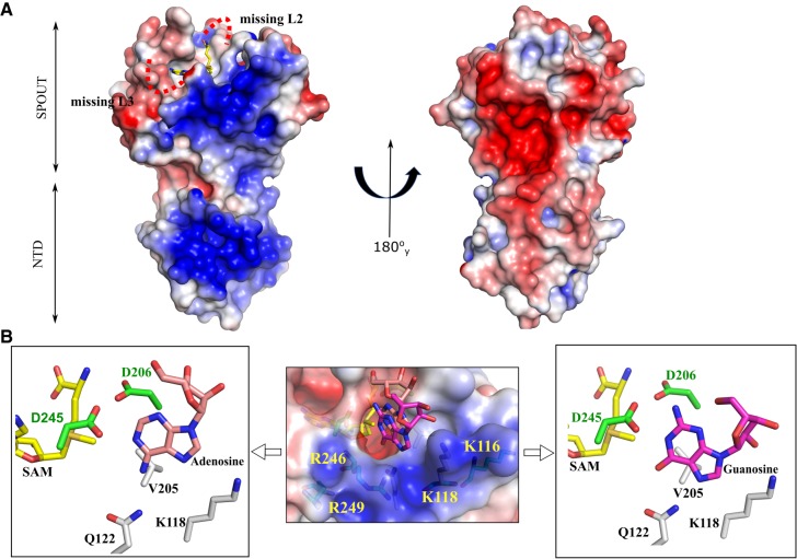
FIGURE 5.
Electrostatic surface potential of TkTrm10 constructs and possible tRNA binding site. (A) Electrostatic potential mapped on the solvent accessible surface of TkTrm10Δ26 in two orientations. The missing loops in the structure are shown by red dotted curved lines in the left panel. For reference, SAM, shown in yellow sticks, is placed in the active site. (B) The middle panel shows the electrostatic potential mapped on the solvent accessible surface of the TkSPOUT_SAM structure with a docked guanosine and adenosine molecule. SAM, guanosine, and adenosine are shown as sticks with carbon atoms colored yellow, magenta, and salmon, respectively. The positively charged residues are labeled on the surface. The left and right panels show a zoomed-in view of, respectively, the docked adenosine and guanosine in the TkSPOUT_SAM active site. The surrounding residues discussed in this study are shown.

