Figure 1.
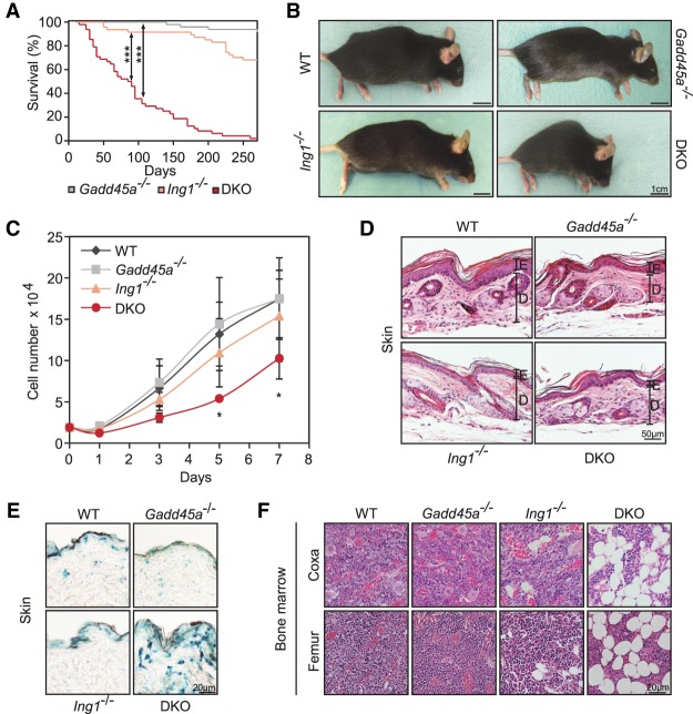
Premature aging of Gadd45a/Ing1 double-knockout mice. (A) Kaplan-Meier survival curves of mice of the indicated genotypes. n = 47–65 mice per genotype. (DKO) Gadd45a/Ing1 double knockout. P-values were based on log-rank test. (***) P < 0.001. (B) Lateral view of mice of the indicated genotypes. Kyphosis is apparent in eight of 42 analyzed double-knockout mice. (C) Growth curves of MEF cells. Total cell numbers were counted at the indicated time points. Data are presented as mean values of three independent MEF lines per genotype ±SD. (*) P < 0.05. (D) Histological image (hematoxylin and eosin [H&E] stain) of hindlimb skin. (E) Epidermis; (D) dermis. n = 1–4 animals per genotype. (E) Representative histological images of SA-β-Gal staining of dorsal skin. n = 5 animals per genotype. (F) Representative histological images (H&E stain) of bone marrow within bones from the iindicated regions. n = 3–5 animals per genotype. Lipid vacuole accumulation of varying degrees was observed in four of five Ing1−/− and double-knockout mice, respectively.

