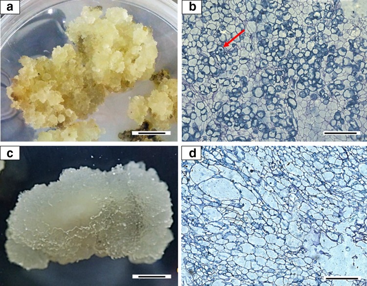
Fig. 1.
Callus developed (morphologically and histologically) from segmented mangosteen young leaves after 120 days of culture on MS medium. a Friable callus on medium supplemented with 4.44 µM BAP and 4.52 µM 2,4-D, showing b the isodiametric cells with dense nucleus (red arrow). c Friable and watery callus on medium supplemented with 4.44 µM BAP and 4.14 µM picloram, showing d cells with large vacuoles. Bars for a and c represents 1 cm; b, d represents 400 µm

