
Figure 2 |. Neural circuitry of efferent and afferent innervation in the kidney.
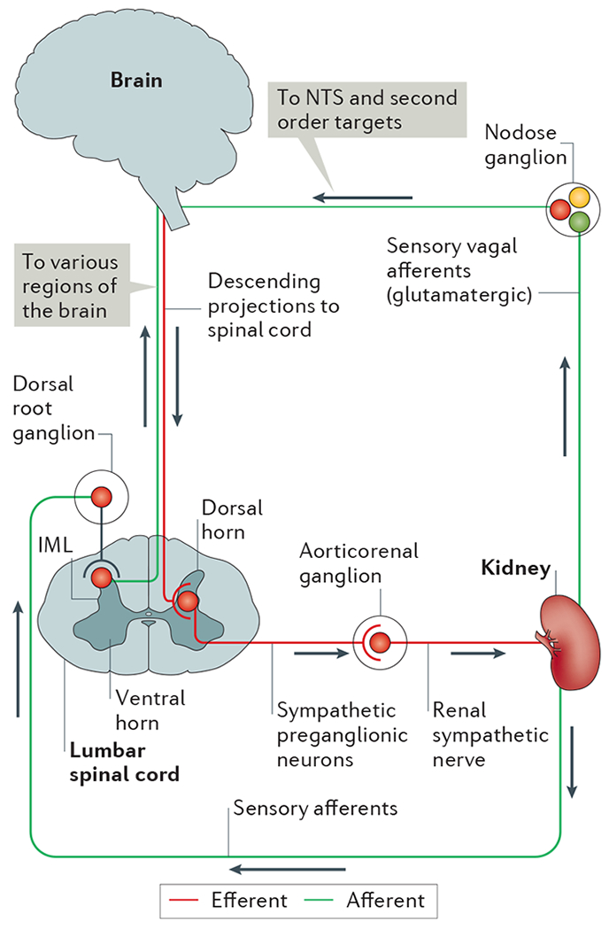
In the efferent pathway, descending projections from the brain (for example, from brainstem and hypothalamic regions) synapse on sympathetic preganglionic neurons in the intermediolateral cell column (IML) of the thoracic and lumbar spinal cord. Sympathetic preganglionic neurons in the lumbar spinal cord project through the ventral horn to the aorticorenal ganglion and activate the renal sympathetic nerve, which innervates the kidney and releases noradrenaline, neuropeptide Y and ATP. Sensory afferents in the kidney, which release substance P and calcitonin gene-related peptide, have their cells bodies in the dorsal root ganglia and synapse centrally on interneurons in the dorsal horn of the spinal cord that transmit information to various regions of the brain. Although the kidney does not seem to have vagal efferent input, some evidence suggests that vagal afferent neurons arising in the nodose ganglion innervate the kidney, thus providing a pathway for integration with the nucleus tractus solitarius (NTS) and subsequent second order neuronal targets in the brain.