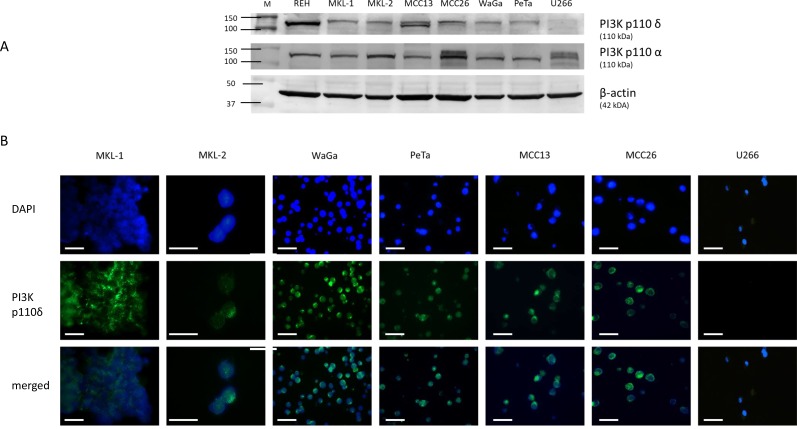
Figure 2.
Assessment of PI3K p110δ expression by Western blotting (A) and immunofluorescence microscopy (B). (A) Expression level of PI3K p110δ and PI3K p110α in cell lysates of REH, MKL-1, MKL-2, WaGa, PeTa, MCC13, MCC26, and U266. All cell lines except U266 (PI3K p110δ negative control) revealed a specific PI3K p110δ protein expression at 110 kDa, whereas PI3K p110α expression was detected in all cell lines. The loading control β-actin is located at 42 kDa. (B) The corresponding IFM photographs for DAPI, PI3K p110δ and merged are shown for these cell lines. The merged photograph identifies PI3K p110δ expression within the cytoplasm. According to Figure 2A U266 cells reveal no detectable PI3K p110δ expression by IFM. The photos were taken with 63× magnification. The scale bars represent a length of 100 µm.

