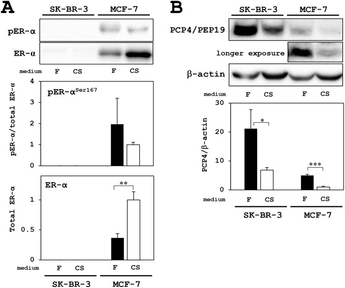
Figure 1. Western blotting analysis for PCP4/PEP19 and ER expression in MCF-7 and SK-BR-3 cells.
(A) ER and active phosphorylated-ER were only detected in MCF-7 cells but not in SK-BR-3 cells. Although total ER-α expression was increased in the culture with charcoal-stripped FBS supplemented medium (CS: open column), the expression levels phosphorylated-ER-α (pER-α) relative to total ER-α were not different in both culture conditions; FBS (F: closed column) and charcoal-stripped FBS (CS: open column) supplementation in the medium. (B) PCP4/PEP19 was highly expressed in SK-BR-3 cells, but not in MCF-7 cells, and the expression was decreased in the culture with CS medium (upper blot). In MCF-7 cells, longer exposure revealed PCP4/PEP19 bands, which were decreased in the culture with CS medium (middle blot). Expression levels of PCP4/PEP19 were normalized by those of β-actin and compared between culture conditions (F: FBS as closed column, CS: charcoal-stripped FBS as open column). * p<0.05, ** p<0.01 and *** p<0.001.

