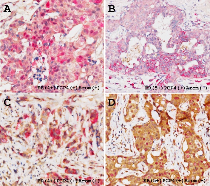
Figure 9. Representative immunohistochemical demonstration of PCP4/PEP19 and aromatase expression in ER-positive human breast cancer tissues.
Thirty four % cases of the breast cancers were positive for both PCP4/PEP19 and aromatase. PCP4/PEP19 positive cells (red) and aromatase positive cells (brown) are mixed in the breast cancer tissues, which results in a mosaic pattern (A and C). Some carcinoma cells express both PCP4/PEP19 and aromatase, having red-brown colored cytoplasm (A, C), while some were only positive for PCP4/PEP19 (B). In some cases, nuclear localization of PCP4/PEP19 was observed in the aromatase-positive breast cancer cells (D).

