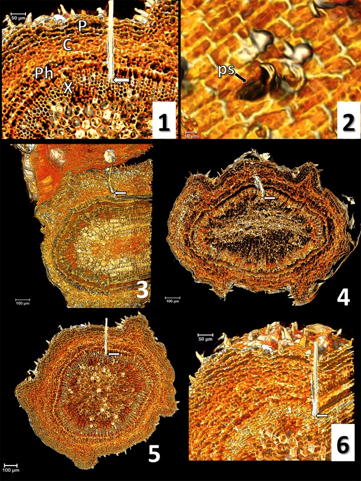
Fig 1. Micro-CT volume rendering reconstruction images and correlation waveform/stylets position.
1) olive anatomy: P (periderm), C (cortex), Ph (phloem), X (xylem) (nomenclature according to Ruiz et al. [45]); 2) probing site (ps); 3) waveform C; 4) waveform Xc; 5) waveform Xi; 6) Xi, lateral view. White arrows indicate the stylets tip.

