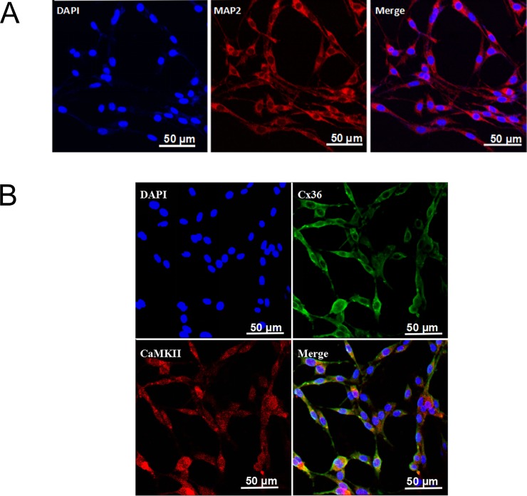
Fig 1. Immunofluorescence staining of MAP2 in PC12 cells and colocalization of Cx36 and CaMKII in PC12 cells.
(A) PC12 cells exhibited neuron-like characteristics. MAP2-positive staining was in red, and the nuclei were stained with DAPI in blue. (B) Colocalization of Cx36 and CaMKII in PC12 cells. The positive Cx36 staining was in green and CaMKII was in red. The nuclei were in blue.

