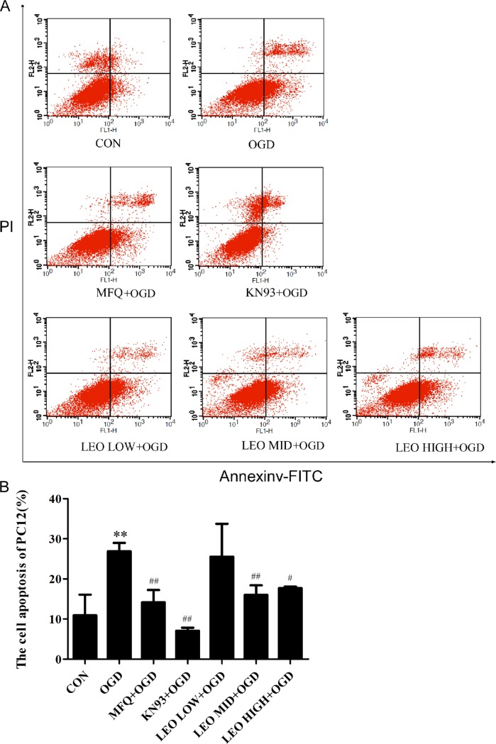
Fig 6. Effect of Leonurine, MFQ,and KN93 on cell apoptosis in OGD-induced PC12 cells.
PC12 cells were incubated under OGD for 2 h, and they were then separately incubated with MFQ (5 μM), KN-93 (2 μM), and leonurine (50, 100, and 200 μg/mL, respectively) for 3 h, followed by culturing under normal conditions for 24 h. Cell apoptosis was detected using Annexin V-FITC/PI staining. (A) Results of flow cytometry. (B) Quantification of cell apoptosis. CON, normal control group; OGD, OGD-for-control group; MFQ+OGD, MFQ-treated group; KN93+OGD, KN93-treated group; LEO LOW+OGD, low-dose leonurine group; LEO MID+OGD, middle-dose leonurine group; LEO HIGH+OGD, high-dose leonurine group. *P < 0.05 compared with the normal control group. **P < 0.01 compared with the normal control group. #P < 0.05 compared with the OGD-for-control group. ##P < 0.01 compared with the OGD-for-control group.

