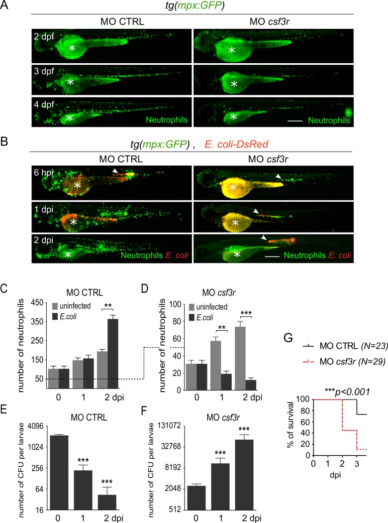
Fig 2. Neutrophils are essential for bacterial clearance.
Tg(mpx:GFP) embryos were injected at the one cell stage with either csf3r morpholino (MO csf3r) to induce neutrophil depletion or a control morpholino (MO CTRL). (A) Steady-state neutrophil populations were imaged repeatedly in individual morphants using GFP fluorescence in both MO csf3r and control conditions between 2 and 4 dpf. (B) Fluorescent E. coli-DsRed were injected in the notochord of csf3r and CTRL morphants. GFP (Neutrophils) and DsRed (E. coli) fluorescence were imaged at indicated time points. E. coli-DsRed (red) disappeared from 1 dpi in control embryos (left panels), while it increased in csf3r morphants at 1 and 2 dpi (white arrowheads) with a concomitant decrease in neutrophil number (green). Scale bars: 400 μm. (C, D) Quantification of total neutrophils in CTRL (C) and csf3r (D) morphants at the indicated time points following PBS (light grey columns) or E. coli (dark grey columns) injections (mean number of cell per larva ± SEM, Mann-Whitney test, two-tailed, **p<0.005, ***p<0.001, Nlarvae = 7–16 per condition, from two independent experiments). (E, F) E. coli log counts (CFU) in CTRL (E) and csf3r morphants (F) (mean number of CFU per larva ± SEM, Mann-Whitney test, two-tailed, ***p<0.001, Nlarvae = 3–4 per condition). (G) Survival curve of MO csf3r and MO CTRL larvae infected with E. coli from 0 to 3 dpi (Nlarvae is indicated in the figure, log rank test, p<0.001, from two independent experiments).

