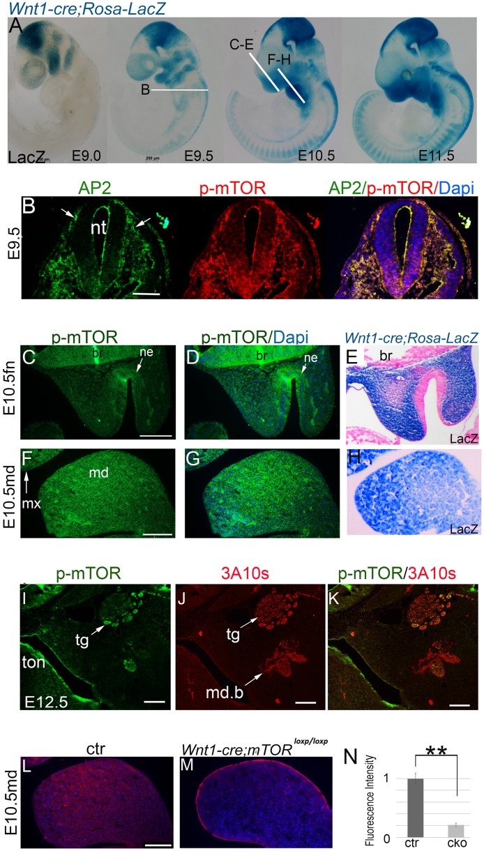
Fig 1. mTOR dynamics during craniofacial development.
(A) Whole mount LacZ staining of Wnt1-cre; Rosa26R embryos. White lines indicate the sectioning planes in relevant images. (B) Immunofluorescence for Ap2-α and p-mTOR in the migrating NCCs at E9.0. (C, D; F, G) Immunofluorescence for p-mTOR in the FN and mandibular prominences at late E10.5. (E, H) Sections of a Lac-Z-stained E10.5 heads, showing NCC distribution in the FN and mandibular prominence. (I, J, K) Co-localization of p-mTOR with a neurofilament marker 3A10. (L, M) Immunofluorescence for p-mTOR in the mandibular prominence of E10.5 mice. (N) Quantification of p-mTOR levels of the mandibular prominences by calculating relative fluorescence intensity, **p<0.01. br: brain; ctr: control; fn: frontonasal prominence; cko: conditional knockout; md: mandibular prominence; md.b: mandibular branch of trigeminal nerve; mx: maxillary prominence; ne: nasal epithelium; nt: neural tube; tg: trigeminal ganglion; ton: tongue. Scale bar in (A): 200 μm; Scale bars in others: 100 μm.

