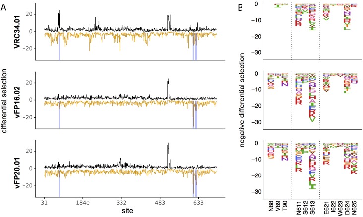
Fig 3. Mutations that better expose the fusion peptide are selected against during antibody treatment.
A. The positive (black) and negative (orange) site differential selection is plotted across the length of the mutagenized portion of Env for each antibody. B. The negative differential selection for regions of interest (regions highlighted in blue in 1A). Left to right, these include the N88 glycosylation motif, the N611 glycosylation, and surface exposed gp41 sites that consistently have mutations depleted upon antibody selection. The height of each amino acid is proportional to the logarithm of the relative depletion of that mutation in the antibody selected condition relative to the non-selected control.

