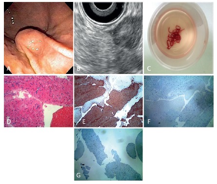
FIGURE 1. A) Gastric subepithelial lesion from the greater curvature of the body; B) linear EUS array demonstrating a lesion from the proper muscle layer; C) EUS-FNA specimens after a total of three needle passes with a 19 gauge needle; D) histopathology confirming a spindle cell tumor (H&E); E) immunohistochemistry stain positive for actin; F) negative for c-kit; G) DOG-1, confirming a gastric leyomioma.

