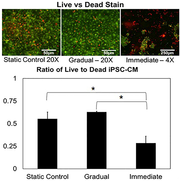
Figure 3:

(Top) Live (green)/dead (red) staining performed on iPSC-CMs following gradual and immediate application of hemodynamic load. The lower magnification image of the immediate sample was required to reliably quantify the increased cell loss experienced within the sample (Bottom) Ratio of live to dead cells shows statistical significance between immediate and other samples (p<0.05, N=3). No statistical significance was found between the static and gradual samples (p>0.05, N=3).
