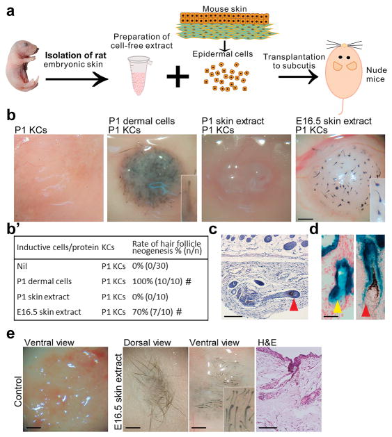
Fig. 1. Cell-free extract from stage-specific embryonic skin induces HF neogenesis.
(a) Preparation of skin extract and HF neogenesis assay. (b, b′) Effect of skin extract on HF induction. Regenerated HFs are pigmented because the P1 keratinocyte preparation also contains melanocyte progenitors. #p < 0.05 with Fisher’s test, compared with P1 KCs only (n = 10). (c) E16.5 skin extract induced new HFs with new DPs (red arrowhead). (d) New HFs formed from lacZ+ keratinocytes exhibited β-galactosidase activity in the epithelium, including the sebaceous gland (yellow arrowhead), but not in the DP (red arrowhead). (e) In full-thickness wounds of nude mice, incorporation of E16.5 extract into dermal equivalents induced HF neogenesis from transplanted C57BL/6 mouse keratinocytes. #p < 0.05 with Fisher’s test, compared with nascent E16.5 skin extract (n = 10). All insets show enlarged images of the regenerated HFs. KC: keratinocyte. Bar: histology, 100 μm; gross images, 500 μm. (For interpretation of the references to colour in this figure legend, the reader is referred to the web version of this article.)

