
Fig. 1.
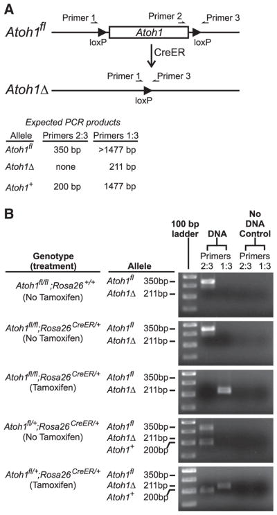
Design of the PCR scheme used to quantify floxed and recombined alleles of Atoh1 following Tamoxifen treatment of Atoh1fl/fl;Rosa26CreER/+ mice. (A) Schematic diagram showing the structure of the floxed Atoh1 allele before (Atoh1fl) and after (Atoh1Δ) Cre-induced recombination of the loxP sites that flank Atoh1. Note that primer 2 is excised following recombination, and the distance between primers 1 and 3 is decreased. The sizes of PCR products expected from the various alleles are indicated. (B) Images of agarose gels of the PCR products from the various genotypes before and after Tamoxifen treatment. Ear punch DNA was used for PCR reactions before Tamoxifen treatment. Isolated epithelial DNA was used after treatment. The floxed Atoh1 alleles were efficiently excised following Tamoxifen treatment.