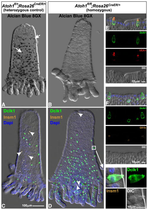
Fig. 19.
Isolated epithelium from adult heterozygous control Atoh1fl/+;Rosa26CreER/+ and homozygous Atoh1fl/fl;Rosa26CreER/+ mice 6 days after initiation of Tamoxifen treatment. (A) The numerous Alcian Blue 8GX positive mucous cells in control epithelium (arrows) were largely absent from Atoh1 deleted (Atoh1fl/fl;Rosa26CreER/+) epithelium (B). (C) Similarly, Insm1+ enteroendocrine cells found in control epithelium (arrowheads) were largely absent from Atoh1 deleted epithelium (D). In contrast, the Dclk1+ Insm1− brush cell population increased dramatically in homozygous epithelium (D) relative to heterozygous control (C). (E, F) The brush cells in Atoh1-deleted epithelium appear normal and stain with the standard brush cell markers including Dclk1, UEA-I, and Gfi1b (see also the enlargement of the region boxed in D).

