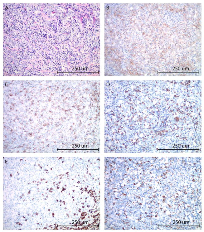
Figure 4.
High grade ovarian carcinoma with marked peritumoral inflammatory cell infiltrate (A). The tumor cells and peritumoral inflammatory cells show moderately to intense membranous staining with PD-L1 immunohistochemistry (B). T-lymphocytes represent the predominant component among the immune cells, highlighted by CD4 (C) and CD8 (D) immunostains. B-lymphocytes (E: CD20 immunostain) and macrophages (F: CD68 immunostain) are present in a smaller proportion. CD56 and TIA immunostains were negative for NK-cells (image not shown). (A: hematoxylin-eosin stain, B: PD-L1 immunostain, C: CD4 immunostain, D: CD8 immunostain, E: CD20 immunostain, F: CD68 immunostain; all images at 200x original magnification).

