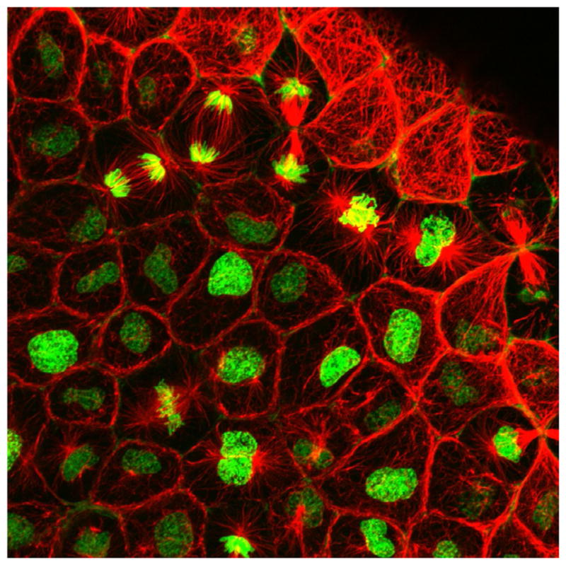
FIG. 1.

A confocal micrograph of an embryonic Xenopus epithelium expressing RNA encoding fluorescent histone H2B (green), and microtubules labeled with immunofluorescence (mouse antitubulin, red). Note the abundance of cells in different stages of mitosis.
