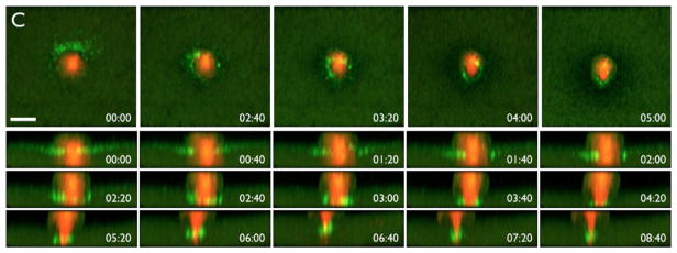
FIG. 2.
Polar body emission in a Xenopus egg expressing fluorescent tubulin (red) to label the meiotic spindle and GFP-rGBD (green) to label active Rho. Top row shows an en face view, revealing formation of a ring-like zone of Rho activity around the nascent polar body; bottom rows show z-views of the same process. Time in min:s. Scale bar is 25μm.
From Bement, W. M., Benink, H. A., & Von Dassow, G. (2005). A microtubule-dependent zone of active RhoA during cleavage plane specification. Journal of Cell Biology, 170(1), 91–101. https://doi.org/10.1083/jcb.200501131.

