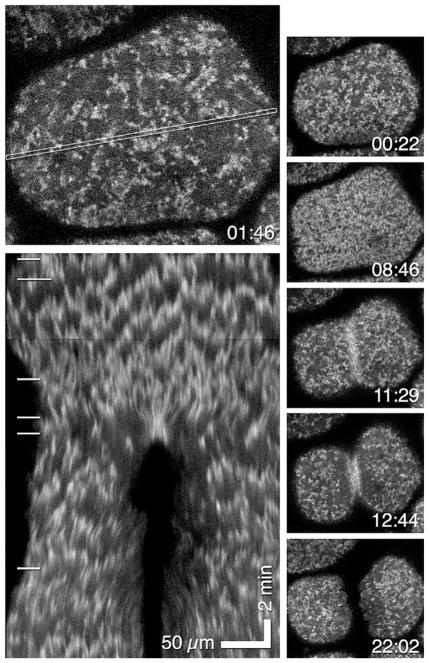
FIG. 6.
Blastomeres from a Xenopus embryo expressing GFP-UtrCh to detect actin filaments. Top left panel shows a blastomere with the area used for the kymograph indicated by a thin rectangle. Bottom left panel shows resultant kymograph which reveals F-actin waves as jagged lines. White lines on left show relationship of kymograph to images above and to the right of the kymograph. Montage shows cortex of blastomere at different times. Cytokinesis ensues at 11:29; this corresponds to fourth white line on kymograph. Time in min:s.
From Bement, W. M., Leda, M., Moe, A. M., Kita, A. M., Larson, M. E., Golding, A. E., et al. (2015). Activator–inhibitor coupling between Rho signalling and actin assembly makes the cell cortex an excitable medium. Nature Cell Biology, 17(11), 1471–1483. https://doi.org/10.1038/ncb3251.

