
Figure 4.
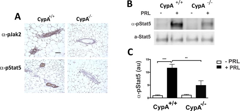
Loss of CypA results in decreased PRL-induced pJak2 and pStat5 at the immunohistochemical (Panel A) and immunoblot (Panels B + C) levels. Thirty minutes following PRL stimulation (10 ug PRL, IP), mouse mammary glands from Cyp+/+ and −/− mice (20-week virgins) were harvested, paraffin-embedded and immunostained with anti-pJak2 or –pStat5 antibodies (Bar = 100 microns) as shown in Panel A. in Panels B + C, tissue explants from CypA+/+ and CypA−/− mice were stimulated for 15 min with PRL, prior to harvest, homogenization, and immunoblot analysis with anti-pStat5 antibodies. Quantitation of pStat5 immunoblot analysis in Panel B is presented in Panel C; n=3 mice, **p < 0.01; ***p < 0.005.