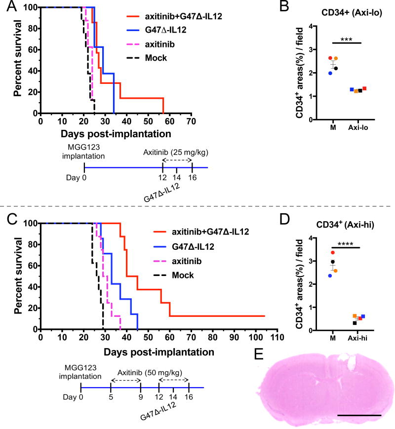
Figure 2. Axitinib treatment in combination with intratumoral G47Δ-mIL12 in athymic mice bearing human MGG123 GSC-derived brain tumors.
A. Kaplan-Meier survival curve. Athymic mice were implanted with MGG123 human GSCs on day 0, axitinib (25 mg/kg) or vehicle solution was injected intraperitoneally from days 12 to 16, and/or G47Δ-mIL12 (5 × 10⁴ pfu) or PBS injected intratumorally on day 14. n=8, except for combination, n=7. Mock vs. axitinib, p=0.16; mock vs. G47Δ-mIL12, p=0.0003; mock vs. combination, p=0.0003; axitinib vs. combination, p=0.0008; G47Δ-mIL12 vs. combination, p=0.78. B. Immunohistochemical staining of CD34+ endothelial cells in brain tumor sections from mice treated with low-dose axitinib (25 mg/kg). Athymic nude mice implanted with MGG123 GSCs (5 × 10⁴) on day 0 and treated with axitinib (25 mg/kg) or vehicle solution following the schema shown in A. Twenty-four hours after the last axitinib injection (day 17), animals were sacrificed and brains collected. Formalin-fixed and paraffin-embedded brain tumor sections were stained for CD34+ endothelial cells. Scatter plot (each animal 1 point) showing the quantification of CD34+ areas (10× objective) from 5 fields / tumor section (2 sections / mouse; n=4 mice/group). Quantification of CD34+ areas was done by ImageJ software (NIH). Counter was blinded to the experiment. C. Kaplan-Meier survival curve. Athymic mice were implanted with MGG123 human GSCs and treated with G47Δ-mIL12 or PBS intratumorally on day 14 (as in A). High dosage of axitinib (50 mg/kg) or vehicle solution was administered intraperitoneally from days 5 to 16 (2 cycles of 5 days on and 2 days off). n=8, except for G47Δ-mIL12, n=7. The long-term surviving mouse from the combination group was sacrificed on day 104, and tumor was not present, shown in E. Mock vs. axitinib, p=0.009; mock vs. G47Δ-mIL12, p=0.002; mock vs. combination, p=<0.0001; axitinib vs. G47Δ-mIL12, p=0.08; axitinib vs. combination, p=<0.0001; G47Δ-mIL12 vs. combination, p=0.02. D. Immunohistochemical staining of CD34+ endothelial cells in brain tumor sections from mice treated with high-dose axitinib (50 mg/kg). Same data as in Fig. 3B. Mean ± SEM. *** P<0.001, **** P<0.0001. E. Hematoxylin and eosin staining of brain section of long-term survivor mouse in C. Mouse was sacrificed on day 104 and H&E stained brain section showing the needle track wound (right hemisphere) with no evidence of tumor. Bar = 0.1 inch.

