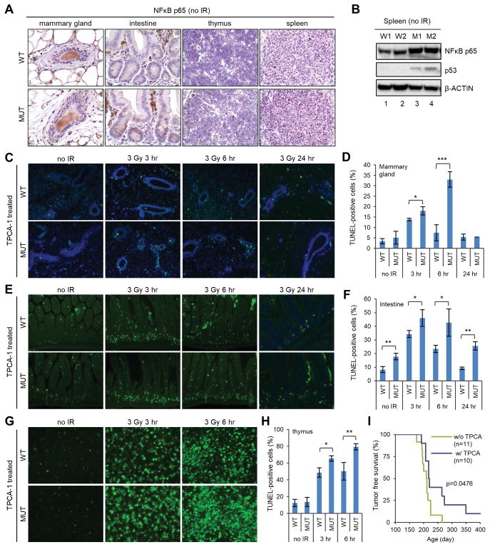
Figure 7.
Evidence for a role of NFκB in apoptosis and tumor development in Palb2 mutant mice after radiation. (A) Representative IHC images of NFκB (p65) in the mammary gland, intestine, thymus and spleen of wt and mutant mice. (B) Western blots showing the amounts of NFκB (p65) and p53 in the spleen of wt and mutant mice. Two pairs of mice (W1, W2 and M1, M2, respectively) were tested. (C,D) Representative TUNEL staining images (C) and quantification of TUNEL-positive cells (D) in the mammary gland of TPCA-1 treated wt and mutant mice before and after 3 Gy of radiation. (E,F) Representative TUNEL staining images (E) and quantification of TUNEL-positive cells (F) in the intestine of TPCA-1 treated mice before and after 3 Gy of radiation. (G,H) Representative TUNEL staining images (G) and quantification of TUNEL-positive cells (H) in the thymus of TPCA-1 treated mice before and after 3 Gy of radiation. For all TUNEL experiments, results shown are means ± SD from 3 independent experiments. *, p<0.05; **, p<0.01. (I) Kaplan-Meier tumor-free survival curves of TPCA-1 untreated and treated Palb2 mutant mice after 3 × 2 Gy of radiation. P value is calculated by log-rank analysis using GraphPad Prism7.

