
Figure 1.
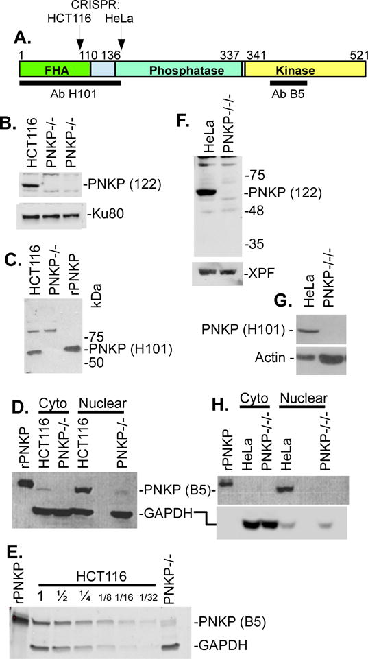
Analysis of PNKP knockout cell lines by western blot. A. Domain structure of PNKP showing location of epitopes recognized by the H101 and B5 anti-PNKP antibodies, and the locations of CRISPR/CAS9-mediated gene knockout. B. Lack of detectable PNKP in whole-cell lysates of PNKP−/− HCT116 cells, as probed with a polyclonal antibody (122) raised against full-length PNKP. Ku80 is a loading control. C. Lack of detectable PNKP in nuclear extracts of same cell line, using the N-terminal H101 antibody. A nonspecific band at ~90 kD serves as a loading control. rPNKP = 50 ng His-tagged recombinant human PNKP. D. Detection of a trace of an apparent truncated PNKP in PNKP−/− HCT116 nuclear extract, using the C-terminal B5 antibody. GAPDH is the loading control. E. Estimation of the level of truncated PNKP in PNKP−/− HCT116 nuclear extract, by comparison with serial dilutions of WT extract. F-H. Lack of detectable PNKP in whole-cell lysates (F, G) or nuclear extract (H) of PNKP−/−/−HeLa cells by any of the three antibodies, with XPF, Actin and GAPDH as loading controls. Nuclear and cytoplasmic fractions were separated using a fractionation kit (BioVision).