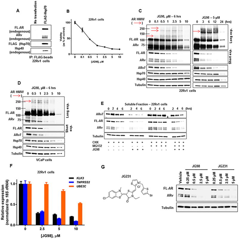
Figure 4.

Pharmacologic targeting of Hsp70 destabilizes the AR/ARv7 signaling axis in CRPC cells.
(A) Lysate from 22Rv1 CRPC cells transfected with FLAG-Hsp70 was probed with FLAG-beads. Bound protein was detected by WB.
(B) Viability of 22Rv1 CRPC cells treated with increasing concentrations of JG98 for 72 hours as determined by MTT assay. Data represent a mean of four to six replicates +/− S.D.
(C) WB of cell lysate from 22Rv1 CRPC cells treated with increasing concentrations of JG98 for 6 hours (left) or 5 μM JG98 for up to 24 hours (right). Red arrows note detection of HMW AR species detected with AR antibody N-20.
(D) WB of cell lysate from VCaP CRPC cells treated with increasing concentrations of JG98 for 6 hours. Red arrow notes detection of HMW AR species detected with AR antibody N-20.
(E) WB of the stability of soluble AR, ARv, and ARv7 from 22Rv1 CRPC cells following pre-incubation with 10 μM MG132 and/or 10 μg/mL cycloheximide (CHX) for 1 hour, followed by +/− 1 μM JG98 for 6 additional hours. Equal amounts of protein (10 μg) were loaded for WB of soluble and insoluble fractions (see also Supplemental Figure 4C).
(F) RT-qPCR of AR/ARv7 target genes, normalized to 18S rRNA, from 22Rv1 CRPC cells treated with increasing concentrations of JG98 for 16 hours. Data represent a mean of three replicates +/− S.D.
(G) Structure of JG231 (left). WB of cell lysate from 22Rv1 CRPC cells treated with increasing concentrations of JG98 or JG231 for 6 hours (right).
Experiments were repeated at least three times.
