
Figure 2. Genetically inhibiting PTP-PEST expression in human GSCs leads to enhanced invasion in vitro.
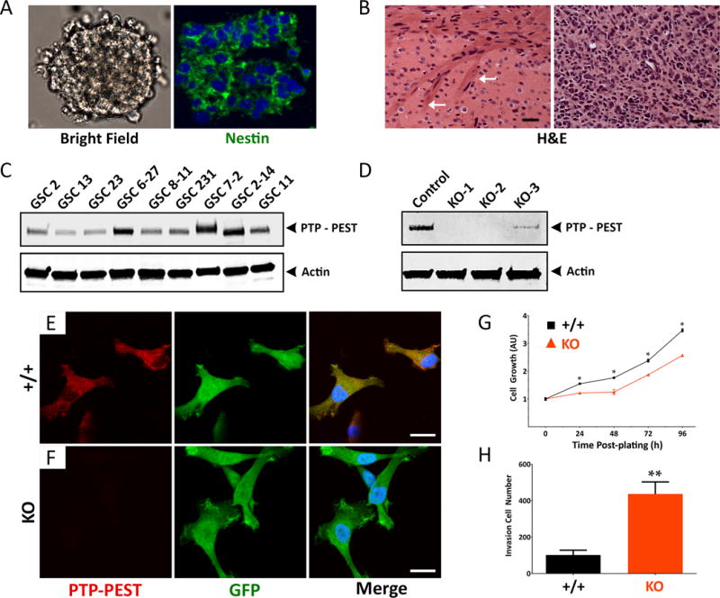
(A, B); Stem-like glioblastoma cells (GSCs) express the neural stem cell protein nestin and grow as free-floating spheroids in serum-free media (A). GSCs generate well-vascularized and invasive tumors after injection into the mouse brain (B). Arrows in the left panel (B) identify invasive GBM cells within white matter of the corpus callosum. Scale bars, 50 μm. (C); Comparative immunoblot showing PTP-PEST protein expression in detergent-soluble lysates from 9 different primary GSC cultures. (D); Anti-PTP-PEST immunoblot confirming CRISPR/Cas9 gene editing strategies to delete PTPN12 in GSC6-27 cells. Controls cell pools were generated with pLentiCRISPR lentivirus expressing GFP and Cas9 without targeting gDNAs, whereas KO cell pools were generated using lentivirus expressing GFP, Cas9 and gDNAs targeting three different regions of PTPN12. (E, F); Adherent control (E) and PTPN12 KO GSC6-27 cells (F) were labeled with anti-PTP-PEST and anti-GFP antibodies and visualized with fluorescent secondary antibodies. Scale bars, 200 μm. (G); Control and PTPN12 KO GSC6-27 spheroids were analyzed for viability over four days in vitro. Note that in comparison to control GSC spheroids, PTPN12 KO-1 spheroids show diminished viability, **p<0.001. Similar results were found with the PTPN12 KO-2 cells. (H); Analysis of invasive capacities of control and PTPN12 KO GSC6-27 cells. PTPN12 KO-2 spheroids were dissociated and three-dimensional cell invasion through Matrigel transwell assays, revealing increased invasion in cells with low levels of PTPN12 gene expression, **p<0.001.