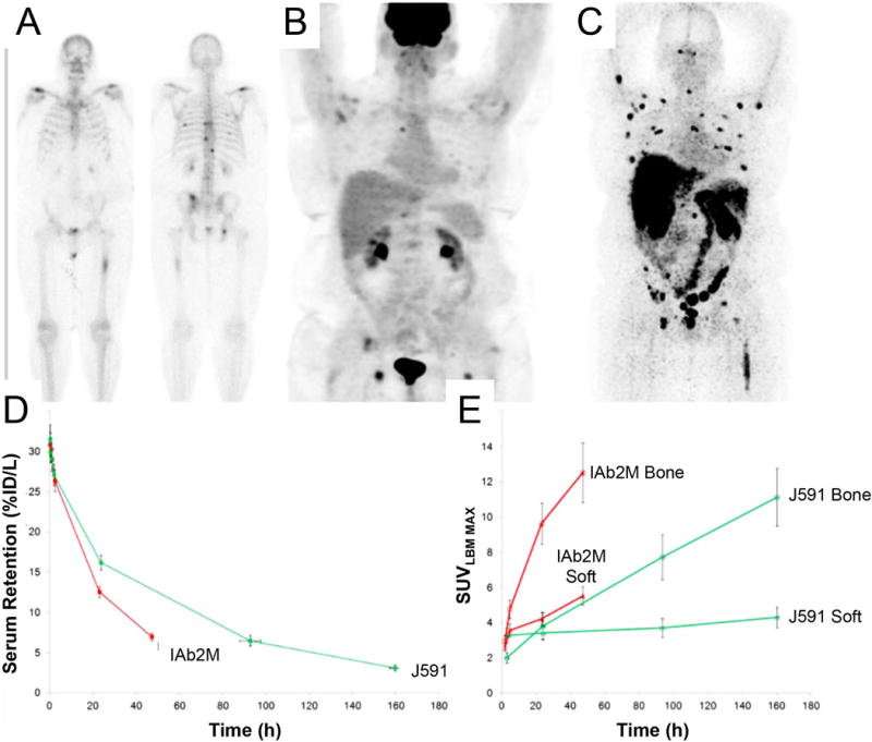
Figure 1. Confirmation of Malignancy.

Differences in lesion detection in a metastatic prostate cancer patient using 99mTc-MDP (bone scan) showed lesions in the ribs and vertebrae A), 18F-FDG PET scan displayed uptake in the femur and in the vertebrae B), and 89Zr-IAB2M imaging identified more true-positive lesions than 99mTc-MDP and 18F-FDG C). Optimization of pharmacokinetics. A comparison of serum clearance D) and lesion uptake E) between 89Zr-IAB2M (minibody) and 89Zr-J591 (full length mAb cognate) over time. This research was originally published at JNM Pandit-Taskar N,Donoghue JA, Ruan S, et al. First-in-Human Imaging with 89Zr-Df- IAB2M Anti-PSMA Minibody in Patients with Metastatic Prostate Cancer: Pharmacokinetics, Biodistribution, Dosimetry, and Lesion Uptake. J Nucl Med.2016;57(12):1858–1864. © by the Society of Nuclear Medicine and Molecular Imaging, Inc. (reprinted with permission from ref.44).
