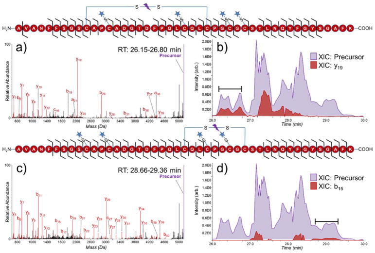
Figure 4.
Deconvoluted UVPD spectra of (a) partially reduced peptide AVANFFSGSCAPCADGTDFPQLCQLCPGCGCSTLNQYFGYSGAFK (3+) (reduced/alkylated disulfide bonds 7 and 8, with intact disulfide C158-C174) and (b) extracted ion chromatograms of the precursor (purple, 5143.16 Da) and diagnostic y19 ion (red, 2248.96 Da) from serotransferrin. Deconvoluted UVPD spectra of (c) partially reduced peptide AVANFFSGSCAPCADGTDFPQLCQLCPGCGCSTLNQYFGYSGAFK (3+) (reduced/alkylated disulfide bonds 6 and 7, with intact disulfide C171-C179) and (d) extracted ion chromatograms of the precursor (purple, 5143.16 Da) and diagnostic b15 ion (red, 1690.59 Da) from serotransferrin from serotransferrin. The spectra in (a) and (c) were averaged over 26.15–26.80 minutes and 28.66–29.36 minutes respectively. Sequence coverage maps of each peptide are shown above their respective spectra/XIC. A star denotes an NEM alkylation site.

