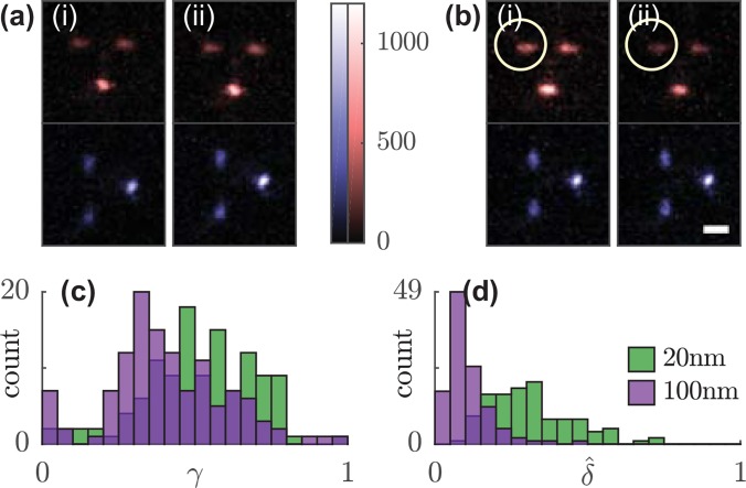
FIG. 2.
Anisotropy and rotation of the orientational second moment vector of fluorescent beads in response to linearly polarized excitation. Sum of 100 Tri-spot PSF images of a single (a) 100-nm and (b) 20-nm bead using (i) x- and (ii) y-polarized excitation. Circle highlights the change in spot brightness. (c) Rotational constraint and (d) normalized rotation in response to x- and y-polarized excitation of 118 fluorescent beads. Green, 20-nm beads; purple, 100-nm beads. Scale bar: 1 μm; color bar: detected photons/pixel.

