
Figure 1: Anatomy of adipose depots.
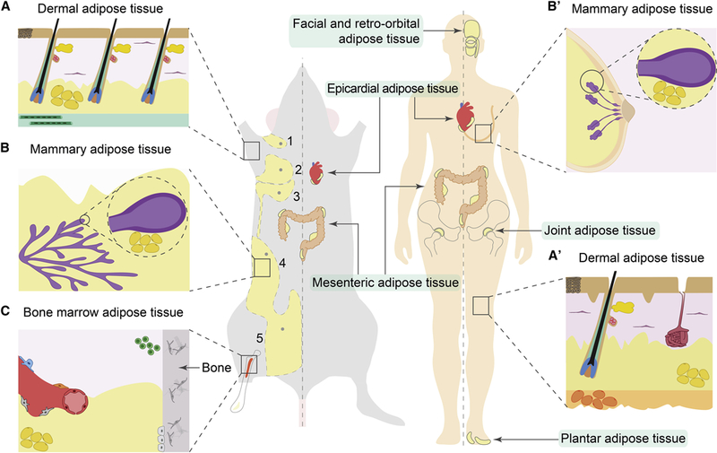
(A, A’) In mouse skin, dermal WAT (dWAT) forms a continuous layer (shown in yellow) separated from subcutaneous WAT (sWAT) by the panniculus carnosus muscle (shown in green). This separation is not prominent in human skin, where dWAT is continuous with underlying sWAT (orange) (A’). Dermal WAT closely associates with HFs and prominently remodels during hair growth cycles. Lower portion of actively growing HFs (shown in red, blue and green) resides within dermal WAT, in close contact with adipocytes. The upper portion of the HF, housing the hair shaft, and containing the HF stem cell compartment and sebaceous gland (shown in red and yellow, respectively) and in humans, a sweat gland (maroon) are also shown. (B, B’) In the mammary gland, adipose tissue (yellow) closely associates with the gland’s epithelium (purple) and undergoes cyclic remodeling during pregnancy, lactation and involution. As shown on D, female mice have five mammary glands, three thoracic (numbers 1, 2 and 3) and two inguinal (numbers 4 and 5), each with its own adipose pads. (C) Specialized adipose tissue is associated with the bone marrow. Micro-anatomically and functionally, it is subdivided into constitutive (yellow) and regulated (red) depots. Adipocytes are shown in yellow, hematopoietic progenitors in green and other mesenchymal bone marrow cell types in grey. Anatomic location of the following additional adipose depots in mouse and human are shown: facial and retro-orbital, epicardial, joint, mesenteric, and plantar adipose tissues.