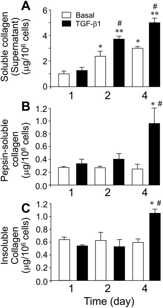
Figure 1.

Time-dependent stimulation of collagen synthesis and deposition in fibroblasts incubated with and without TGF-β1. (A) Soluble collagen in the supernatant. (B) Pepsin-solubilized collagen fraction associated to cell monolayer. (C) Insoluble collagen deposited into the matrix. Collagen fractions were determined from cells incubated for 1 to 4 days in the absence (white bars) or presence of 5 ng/ml TGF-β1 (black bars) as described under Materials and Methods. Values are represented as μg collagen per million of cells (mean ± SEM, n = 6; *P < 0.05 or **P < 0.01vs one day in the absence of TGF-β1, and #P < 0.05 vs the corresponding time value in the absence of TGF-β1).
