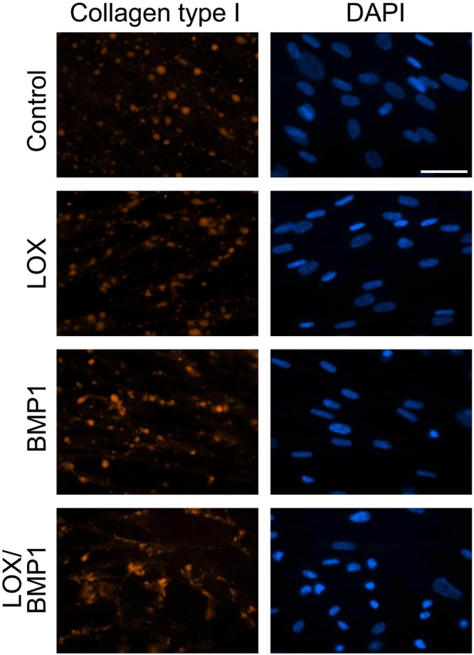
Figure 5.

Immunofluorescence analysis of collagen type I deposition from fibroblast cultures exposed to LOX/BMP1 supernatants. Fibroblasts exposed to control or LOX/BMP1 supernatants and incubated in the presence of TGF-β1 for 4 days were processed for immunofluorescence analysis of collagen type I as described under Materials and Methods. Micrographs shown correspond to representative results of staining for collagen type I (red) and nuclei using DAPI (blue) performed twice with two independent preparations. Bars = 50 μm.
