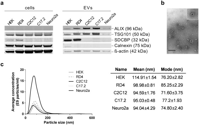
Figure 1.
Characterization of extracellular vesicles (EVs). (a–c) EVs were characterized by Western blotting (5 × 10⁹ particles loaded per well) (a), electron microscopy (HEK, scale bar = 500 nm) (b) and Nanoparticle Tracking Analysis (NTA) (c), confirming the presence of ALIX, TSG101, SDCBP (syntenin, human reactive antibody) and cup-shaped morphology of particles. ß-actin serves as a loading control for cell samples. Full-length western blots can be found in Supplementary Figure S1. Mean/mode size (nm) ± SEM for NTA measurements are depicted.

