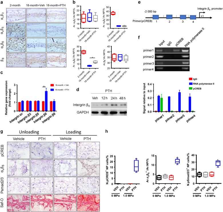
Fig. 4.
PTH induces integrin αvβ6 expression to activate latent TGF-β. a Immunostaining images showing various types of integrin expressions in IVD tissue from 18-month-ld mice injected with PTH or vehicle and quantitative analysis (b). Scale bar, 50 μm. c qRT-PCR analysis of the mRNA levels of various integrin in NP tissue from 18-month-old mice injected with PTH or vehicle. Results reported as fold change. d Western blot analysis of integrin β6 expression in NP cells of 18-month-old mice at different time points post PTH injection (PTH1-34, 100 nmol·L-1). e, f Chromatin immunoprecipitation assay with four different potential pCREB binding sites (primers 1, 2, 3 and 4) in the β6 integrin promoter. g pCREB, Integrin αVβ6, pSmad2/3, or Safranin-O staining of IVD sections from an IVD ex vivo compression model of 30-month-old rat with treatment of either vehicle or PTH (PTH1-34, 100 nmol·L-1). Scale bar, 20 μm. h Quantitative analysis of the percentage of pCREB, pSmad2/3 positive cells and the Integrin αVβ6 positive areas as a percentage of total IVD area (Ar) of (g). All data are reported as the mean ± s.d. *P < 0.05. n = 8 per group. Statistical significance was determined by one-way ANOVA and Student's t-test

