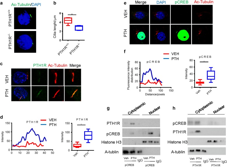
Fig. 7.
PTH stimulates transport of PTH1R to primary cilia of NP cells. a Immunostaining for acetylated α-tubulin (green) and DAPI (blue) showing the length of primary cilia of NP cells from PTH1R-deficient mice or PTH1R+/+ mice. b Quantitative measurements of primary cilia length of (a). c Immunostaining for acetylated α-tubulin or PTH1R showing that PTH-stimulated transport of PTH1R to primary cilia of NP cells. d Quantitative analysis of PTH1R intensity in cilia of (c). e Immunostaining for DAPI, pCREB, or acetylated α-tubulin showing that PTH-stimulated phosphorylation of CREB at primary cilia of NP cells. f Quantitative analysis of pCREB intensity in cilia of (e). g, h Co-immunoprecipitation of cell lysates from NP cells treated with PTH or vehicle using antibody against pCREB and blotted with PTH1R (g) or using antibody against PTH1R and blotted with pCREB (h) showing the interaction between PTH1R and pCREB in the acetylated α-tubulin extracts. *P < 0.05. n = 8 per group. Statistical significance was determined by one-way ANOVA and Student's t-test. All data are reported as the mean ± s.d.

