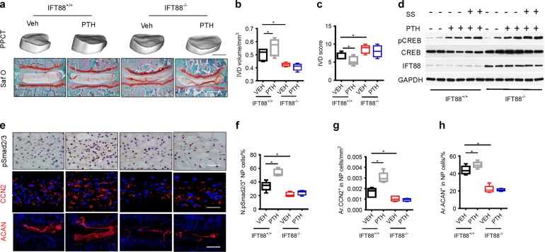
Fig. 9.
Primary cilia regulates PTH signaling in NP cells for disc anabolic activity. a 3D PPCT and immunostaining images showing that PTH effect on IVDs diminished in IFT88−/− mice. PPCT Scale bar, 500 μm. Safranin-O Scale bars, 100 μm. b Quantitative analysis of IVD volume and (c) IVD histological scores of (a). d Western blot analysis of pCREB in NP cells isolated from IFT88−/− or IFT88+/+ mice injected with iPTH or vehicle with or without shear stress. e Immunostaining for pSmad2/3 (brown), CCN2 and ACAN (red) with DAPI (blue) in 2-month IFT88−/− or IFT88+/+ mice treated with PTH or vehicle. pSmad2/3 and CCN2 Scale bar, 50 μm; ACAN Scale bar, 100 μm. f, g and h Quantitative analysis of percentages of pSmad2/3 positive cells in NP area and CCN2 and ACAN-positive area of total IVD area. *P < 0.05. n = 8, per group. Statistical significance was determined by one-way ANOVA and Student's t-test. All data are reported as the mean ± s.d.

