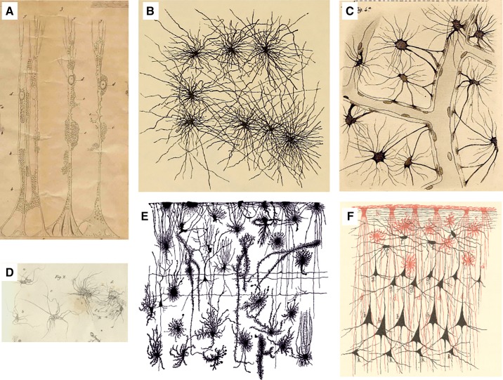
FIGURE 2.
Historic images of astrocytes. A: Müller glial cell of the sheep retina drawn by Max Schulze using a microscope from Amici. y, Brushlike fibrils extending from the outer Müller fiber in the outer granular layer; x, internal limiting membrane; a, opening in the limiting membrane; b, very delicate network of fenestrated membranes similar in the ganglion cell layer; c, network in the so-called molecular layer; d, nuclei as part of the Müller fibers; ee, cavity in which the nuclei or the cells of the internal granular layer are located. [From Schulze (1579). Image has been kindly provided by Prof. Helmut Kettenmann, Max Delbruck Centre for Molecular Medicine, Berlin.] B: cortical astrocytes drawn by Albert von Kolliker (894). C: Camillo Golgi’s drawings of astrocytes contacting blood vessels (583). D: the “Spinnenzellen” of Moritz Jastrowitz (787). E: morphological diversity of neuroglia in human fetal cortex (1458). F: close interactions between neuroglial (red; both interlaminar and protoplasmic astrocytes are clearly presented) and neuronal (black) networks in human brain. [From Schleich (1565).]

