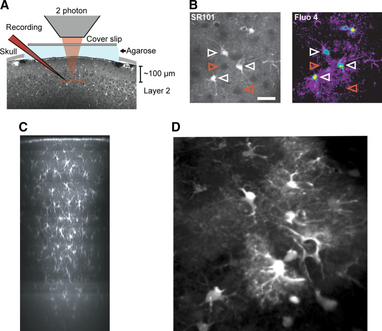
FIGURE 7.
Two photon imaging of astrocytes in vivo. A: experimental setup. Exposed somatosensory cortex was loaded with the specific astrocyte marker sulforhodamine 101 (SR101) and Ca2+ indicator fluo 4-AM. Coverslip and 1% agarose were mounted on top of cranial window to minimize brain pulsation. Recording electrode was loaded with Texas red-dextran (red) and inserted into cortical layer 2 (100–150 μm below pial surface). B: example images showing cortical layer 2 astrocytes double labeled with SR101 and Fluo 4-AM. Only SR101-positive astrocytes also labeled with Fluo 4-AM (white arrowhead). Neurons appeared as dark round shape area (red arrowhead). Scale bar, 30 μm. [A and B from Tian et al. (1750).] C: overview side projection of an SR101-stained area (revealing astrocytes) in mouse neocortex ~30 min after dye application. The image is a maximum-intensity side-projection from a stack of fluorescence images taken through cranial window on an anesthetized mouse. [C from Nimmerjahn et al. (1224). Reprinted by permission from Macmillan Publishers Inc.] D: cortical astrocytes loaded with SR101 and imaged (using two photon confocal system) through the cranial window on an anesthetized mouse. (Image kindly provided by Dr. Hajime Hirose, RIKEN, Japan.)

