FIGURE 9.
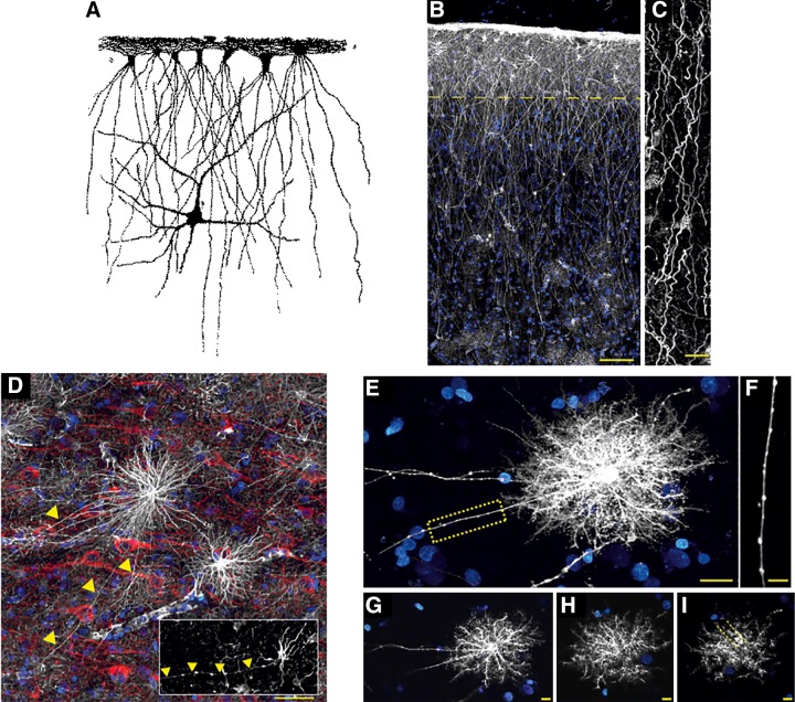
Interlaminar and varicose projection astrocytes in human cortex. A: interlaminar astrocytes as seen by William Lloyd Andriezen (44). B: pial surface and layers 1–2 of human cortex. GFAP staining is in white; DAPI is in blue. Scale bar, 100 μm. Yellow line indicates border between layer 1 and 2. C: interlaminar astrocyte processes. Scale bar, 10 μm. [B and C from Oberheim et al. (1247), with permission of Springer.] D: varicose projection astrocytes reside in layers 5–6 and extend long processes characterized by evenly spaced varicosities. Inset: varicose projection astrocyte from chimpanzee cortex. GFAP, white; MAP2, red; DAPI, blue. Yellow arrowheads indicate varicose projections. Scale bar, 50 μm. E: diolistic labeling (white) of a varicose projection astrocyte whose long process terminates in the neuropil. Sytox, blue. Scale bar, 20 μm. F: high-power image of the yellow box in B, highlighting the varicosities seen along the processes. Scale bar, 10 μm. G–I: individual z-sections of the astrocyte in E, demonstrating long processes, straighter fine processes, and association with the vasculature. [D–I from Oberheim et al. (1248).]

