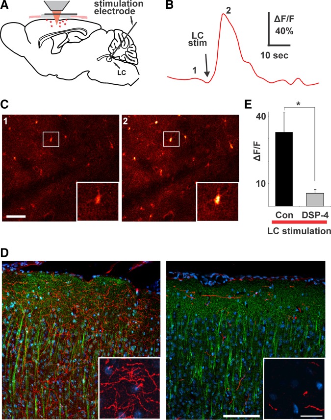
FIGURE 16.
Direct stimulation of locus coeruleus elicits Ca2+ transients in cortical astrocytes. A: the scheme of experimental setup. B: average fluorescence of the image field in C over time showing Ca2+ response to locus coeruleus stimulation. C: fluo 4-labeled astrocytes taken at time points indicated by numbers in B. Insets are blown-up view of boxed area in pictures. Scale bar, 50 μm. D: images from coronal sections through somatosensory cortex illustrating the effect of N-(2-chloroethyl)-N-ethyl-2-bromobenzylamine (DSP-4) on locus coeruleus projection neurons as labeled by antibodies to TH (in red) and MAP-2 (in green) with DAPI labeling of nuclei for orientation. Scale bar, 100 μm (20 μm in inset). E: consistent with the significant reduction in locus coeruleus projection neurons and despite almost twice the stimulation intensity (216 ± 33 vs. 400 ± 39 μA; P = 0.004, t-test), there is a significant reduction in the cortical Ca2+ response to locus coeruleus stimulation in DSP-4-treated animals (P = 0.013, t-test). [From Bekar et al. (123).]

